Fig. 3.

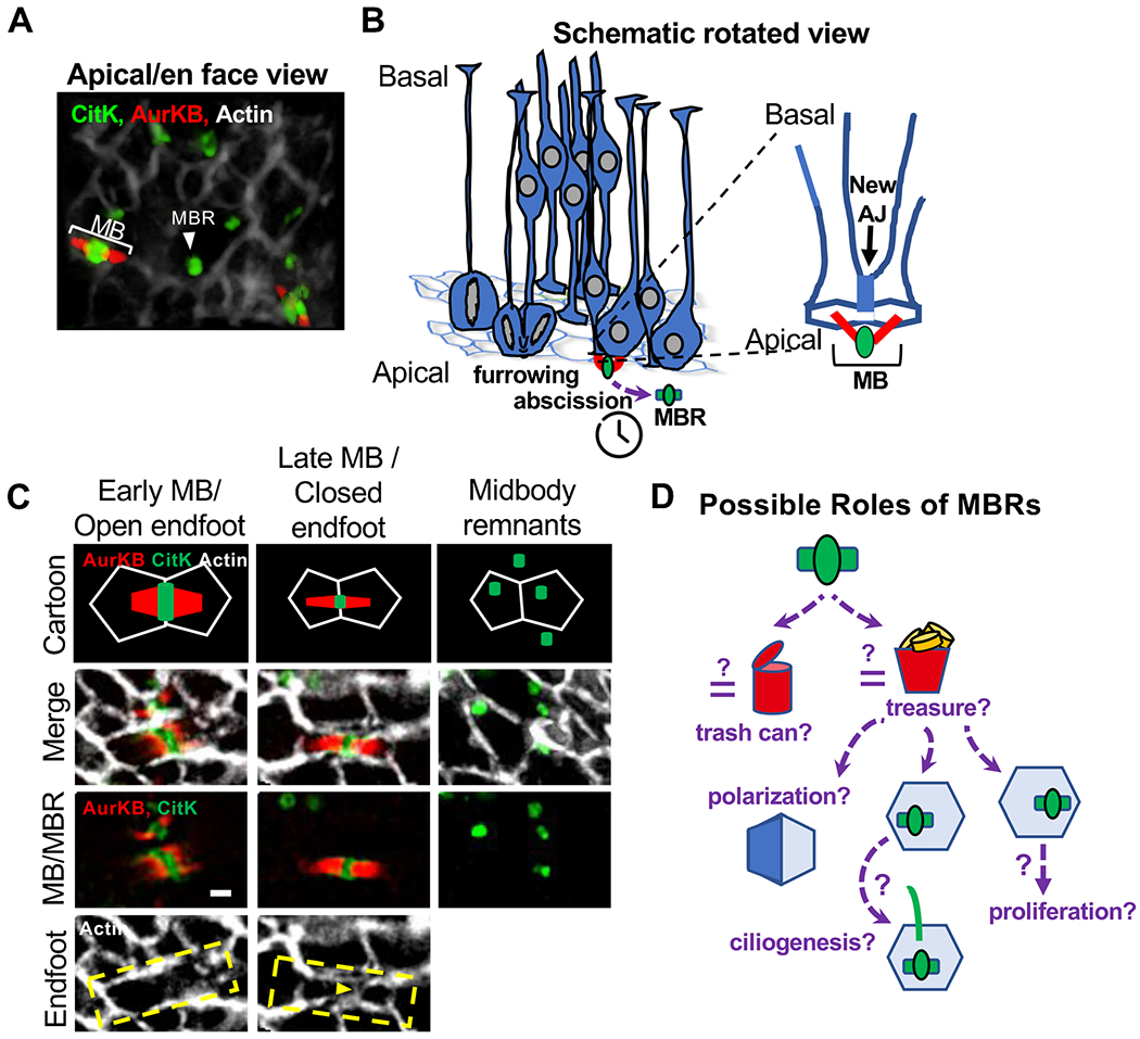
NSC cytokinetic abscission is coordinated with apical membrane segregation and signaling events. (A) En face view of E11 apical membrane where NSC midbodies (MB) form and midbody remnants (MBRs) are deposited. Cortical slab is labeled with phalloidin (actin, apical junctions, AJ), AurKB (MB flanks), and citron kinase, CitK (central bulge and MBRs). (B) Schematic of NSC cytokinesis at the apical membrane. Zoomed view of a pair of daughter cells connected by a midbody and newly forming AJ. (C) MB maturation coordinates with apical endfoot cleavage and new AJ formation. Early midbodies are wider and surrounded by the “open” NSC endfoot. Late midbody is thinner and the endfoot is “closed,” split in two by a new junction (yellow arrowhead) forming between the daughter cells, basal to the MB. MBRs are released at the apical membrane after abscission of both midbody flanks. Scale bar 1 μm. (D) Schematic of proposed roles of MBRs. The MBR could function as a “trash can” to remove unwanted proteins from the newborn daughter cells, or it could be a “treasure chest” for inducing polarization or promoting ciliogenesis or proliferation. See [65••] for methods.
