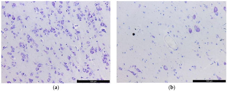
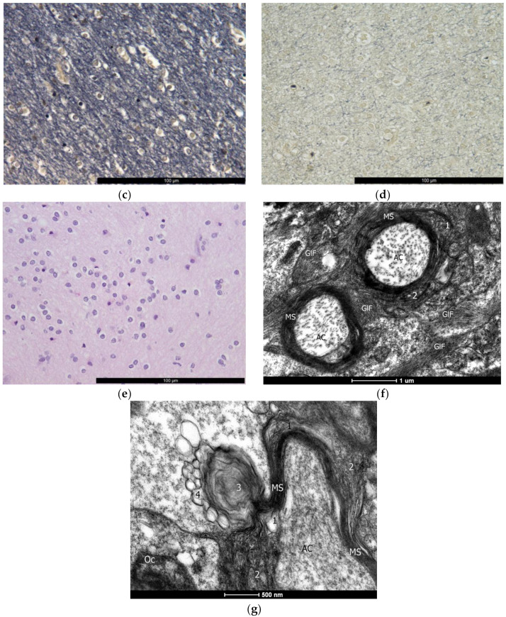
Figure 1.
Structural changes in the epileptic focus in the temporal lobe. Light microscopy: (a) neurons in the cerebral cortex in the comparison group; (b) loss of neurons in the cerebral cortex in a patient with DRE (Nissl staining, 200×); (c) white matter rich in myelin in the comparison group; (d) white matter demyelination in a patient with DRE (Spielmeier staining, 200×); (e) white matter, gliosis (HE, 400×). TEM: (f) destruction of myelin sheaths; bar, 1 μm; 16,500×; (g) destruction of myelin sheaths; bar, 500 nm; 26,500×. 1, areas of lamella rupture; 2, delamination of the sheath; 3, myelin dissociation; 4, vesicular disintegration; 5, grainy myelin disintegration; GlF, gliofibrils; Oc, oligodendrocyte; AC, axial cylinder, axon; MS, myelin sheath.


