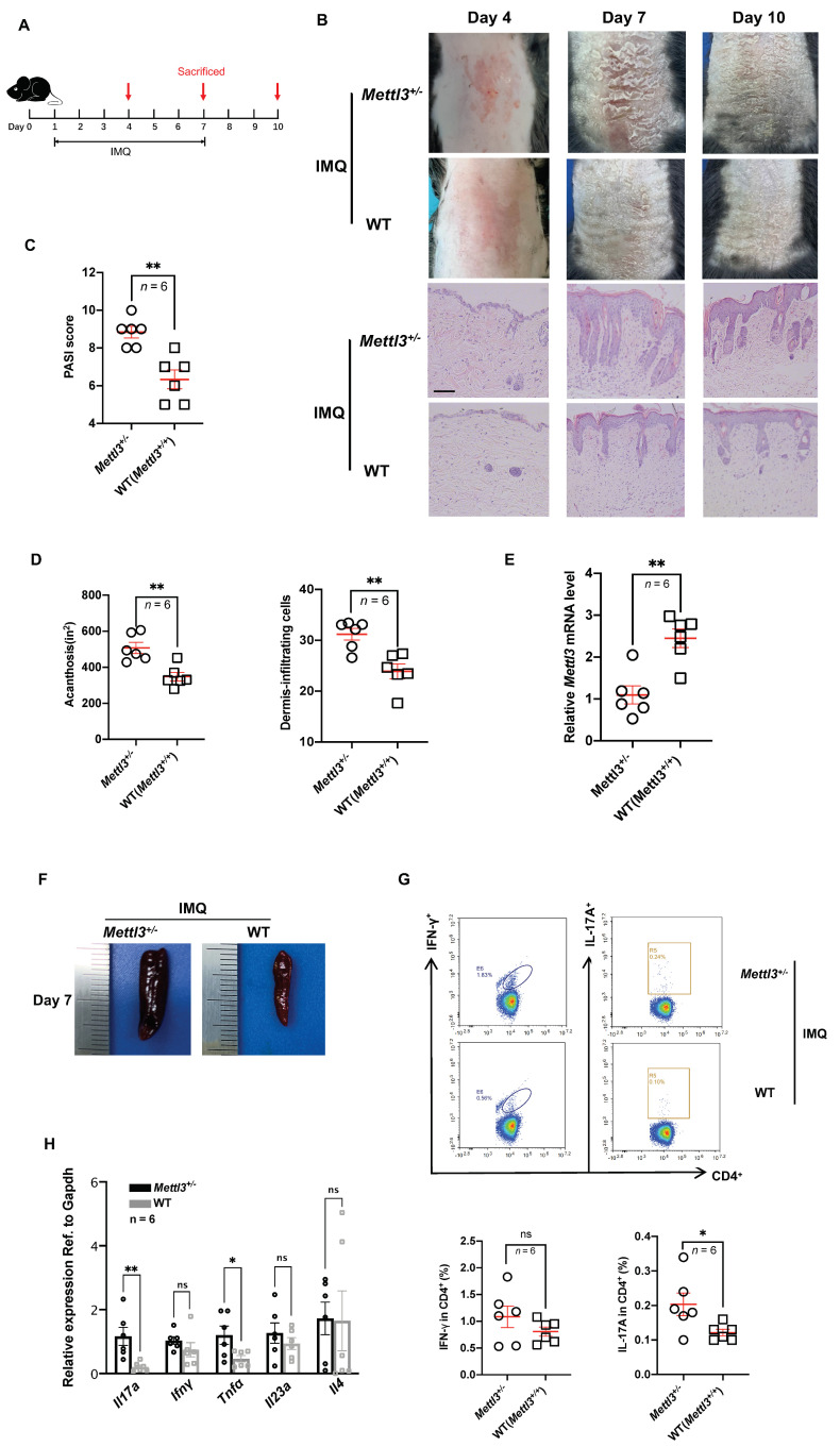
Figure 2.
Heterozygous knockout of Mettl3 accelerated the development of psoriasis. (A) Schematic diagram of the experimental plan on IMQ−treated mice (C57BL/6J). Six mice in each group were sacrificed on days 4, 7, and 10 to conduct experiments. (B) Phenotypic presentation and H&E staining of lesional skin from Mettl3+/− and WT (Mettl3+/+) mice. Scale bars: 100 μm. (C) PASI scores of mice in Mettl3+/− and WT (Mettl3+/+) groups (n = 6). (D) Acanthosis and dermal cellular infiltrates were quantified for Mettl3+/− mice (n = 6) or WT (Mettl3+/+) mice (n = 6). For all measurements in D, the median number of specifically stained dermal nucleated cells was counted in six areas in three sections of each sample. (E) Expression of Mettl3 in skin samples from Mettl3+/− mice (n = 6) and WT (Mettl3+/+) mice (n = 6). (F) The size of spleens of mice in the Mettl3+/− and WT (Mettl3+/+) groups. (G) Representative flow cytometric analysis of Th1 and Th17 cells in splenic CD4+ T cells from Mettl3+/− (n = 6) or WT (Mettl3+/+) (n = 6) mice treated with IMQ for 7 days. (H) The mRNA levels of Il17a, Ifnγ, Tnfα, Il23a, and Il4 in skin samples from Mettl3+/− (n = 6) or WT (Mettl3+/+) mice (n = 6). Data (C−H) were obtained on the seventh day. Data represent the mean ± SEM. * p < 0.05, ** p < 0.01. NS, not significant. Circle, point, triangle and square represent each sample. Two-tailed unpaired Student’s t test (C−E,G,H) was used.

