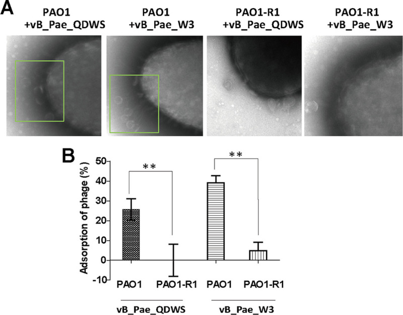
FIG 3.

Phage adsorption assay. (A) PAO1 and PAO1-R1 strains were mixed with phages vB_Pae_QDWS and vB_Pae_W3 at an MOI of 100 for 5 min and subjected to TEM analysis. (B) Adsorption assays of phages vB_Pae_QDWS and vB_Pae_W3 for PAO1 and PAO1-R1 strains. The values were the averages of three measures with standard deviation. T tests were performed to calculate the P-values, and double asterisks indicate statistically significant difference (**, P < 0.01).
