Figure 1.
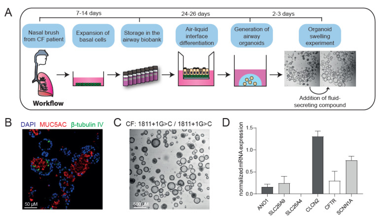
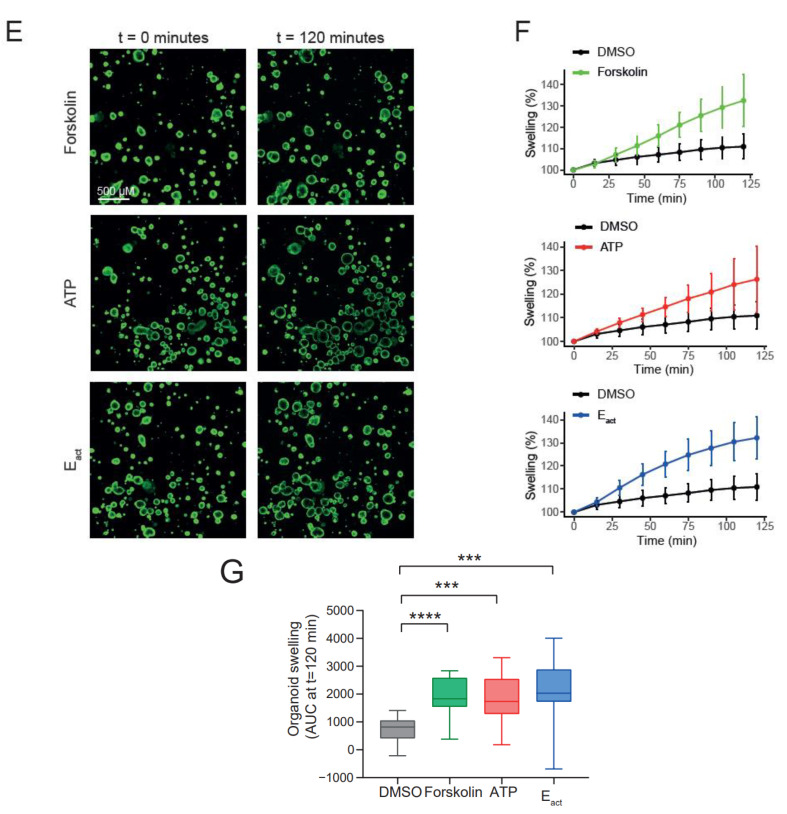
Characterization of CF nasal organoids and CFTR-independent organoid swelling. (A) Schematic representation of the project workflow from nasal brush towards organoid swelling experiments; (B) immunofluorescence staining of nasal organoids with the secretory cell marker MUC5AC (red), ciliated cell marker β-tubulin IV (green) and DAPI (blue) from a CF donor (F508del/S1251N); (C) brightfield image showing intrinsic lumen formation of unstimulated nasal organoids from a CFTR-null donor (1811+1G>C/1811+1G>C); (D) mRNA expression in CFTR-null nasal organoids of the following ion channels/transporters: ANO1 (TMEM16A), SLC26A9, SLC26A4, CLCN2, SCNN1A and CFTR (n = 3 independent donors; W1282X/1717-1G>A, R553X/R553X, G542X/CFTRdele2.3 (21 kb)); (E) confocal images of CFTR-null (G542X/CFTRdele2.3(21 kb)) nasal organoids, stimulated with forskolin (5 µM), ATP (100 µM) or Eact (10 µM) at 0 and 120 min; (F) quantification of CFTR-null (G542X/CFTRdele2.3(21 kb), n = 5 replicates) nasal organoid swelling after stimulation with forskolin, ATP or Eact; (G) area under the curve (AUC) plots of nasal organoid swelling in three CFTR-null donors (n = 3 independent donors; W1282X/1717-1G>A, R553X/R553X, G542X/CFTRdele2.3 (21 kb); 2–6 replicates per donor) after stimulation with forskolin, ATP or Eact. Analysis of difference with control was determined with a one-way ANOVA with Dunnett’s post hoc test (G). *** p < 0.001, **** p < 0.0001.


