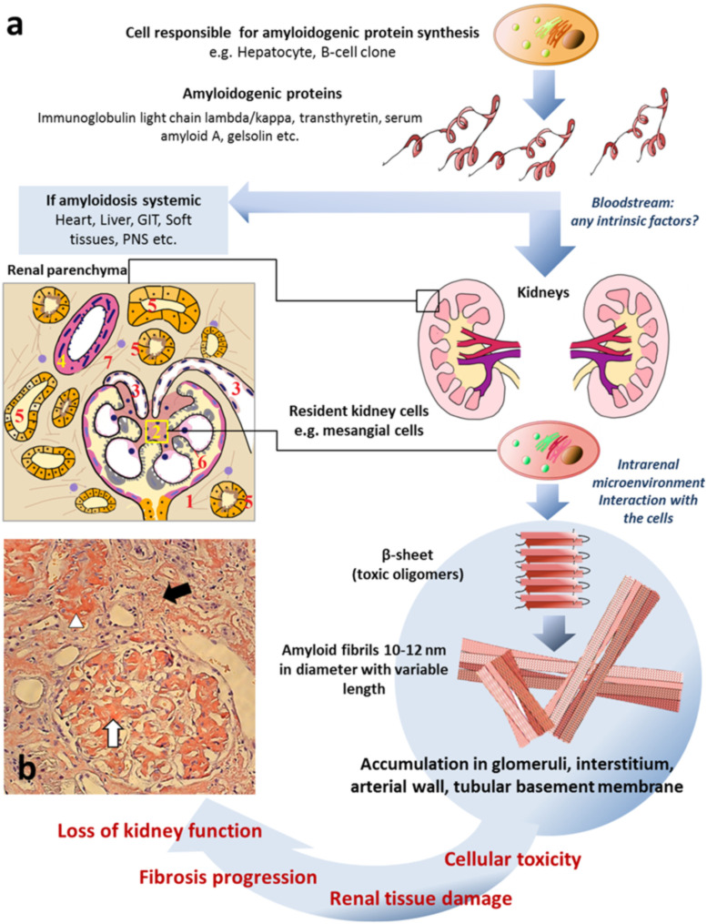
Figure 1.
Common mechanisms of renal amyloidosis. (a) Schematic representation of process of renal amyloidosis formation. GIT—gastrointestinal tract; PNS—peripheral nervous system; scheme of renal parenchyma: 1—glomerulus; 2—mesangium; 3—arterioles; 4—arterial wall; 5—tubules; 6—glomerular basement membrane; 7—interstitium. (b) The microphotograph demonstrating amyloid deposition in mesangium, capillary basement membranes and arterioles of glomerulus (white arrow), interstitium (black arrow) and arterial wall (arrowhead) presented as homogenous Congo-positive masses. Congo red stain, original magnification ×200 (the microphotograph was obtained by V.G. Sipovsky.

