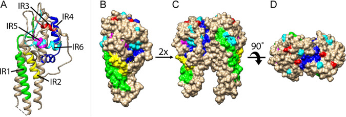
FIG 4.
IR-highlighted three-dimensional structures of VlsE. Structure diagrams of VlsE protein from B31 were prepared in Chimera (Version 1.15) (77) based on the PDB file (accession 1L8W) (19). IRs were highlighted in different colors (IR1 in green, IR2 in yellow, IR3 in red, IR4 in dark blue, IR5 in magenta, and IR6 in cyan). (A) Ribbon diagram showing that IRs tend to form alpha helices. (B) Surface-filled diagram showing membrane surface exposure of IRs in monomeric form. (C) and (D) Dimerized structure models. The structures were oriented to show the membrane-proximal part at the bottom (A, B, and C).

