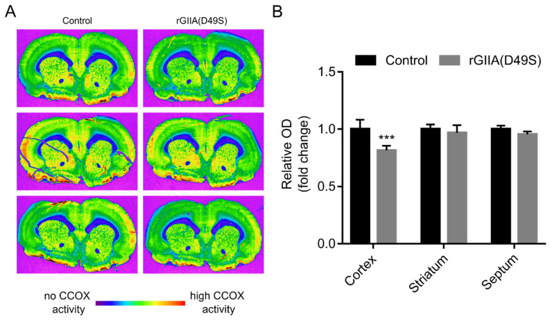
Figure 6.
rGIIA(D49S) also affects the enzyme activity of CCOX ex vivo in tissue sections of rat brain. (A) Rats were sacrificed and coronal cryostat sections (10 µm) were cut through their rostral hippocampus. Consecutive sections were histochemically stained for CCOX activity in the presence or absence (control) of 10 µM rGIIA(D49S) and were imaged using a sensitive black-and-white video camera (see Materials and Methods section for details). Different shades of grey corresponding to different CCOX activity levels were visualized in the pseudo-color spectrum, using MCID M4 image-analysis software. Magnificaation 1.4×. (B) Densitometric analysis of the CCOX staining on cryosections from (A) in different ROIs: cerebral cortex, striatum and septum. Results are presented as means ± SD of fold change in relative optical density (ROD) compared to ROD of untreated sections from three independent experiments, and results that were statistically significantly different from the control are indicated (*** p < 0.001; two-way ANOVA with Bonferroni’s post-hoc test).

