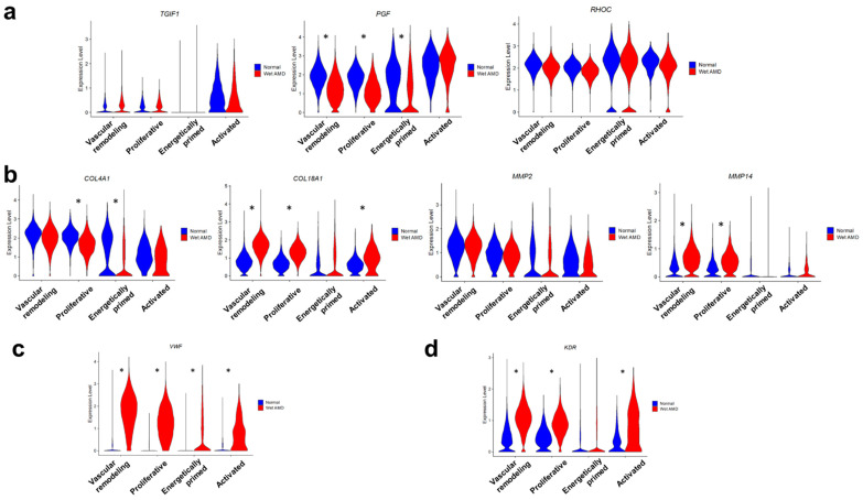
Figure 3.
Benchmarking of human sprouting BOECs to in vivo choroidal neovascularization profiles. (a) Violin plots showing expressions of curated CNV tip endothelial cell markers based on Rohlenova et al. (b) Violin plots showing expressions of collagen genes and matrix metalloproteinases. (c) Violin plot showing expression of VWF, an injury marker, in normal and wet AMD sprouting BOECs. (d) Violin plot showing expression of KDR, a VEGF receptor, in normal and wet AMD sprouting BOECs. Asterisks mark significant differential expression between wet AMD and normal BOECs within each cluster, as determined using FindMarkers.

