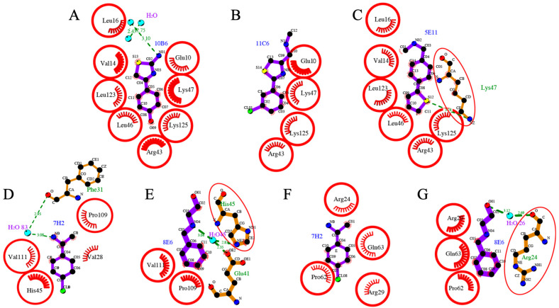
Figure 4.
Molecular interactions of fragment hits binding to SARS-CoV-2 nsp110-126. Hydrophobic interactions are displayed by red half-moons and identical residues are shown with a red circle. Hydrogen-bond interactions are shown with a dotted green line. Three fragment hits (A) 10B6, (B) 11C6 and (C) 5E11 interacting with the residues of SARS-CoV-2 nsp110-126 in binding pocket I. The binding conformation is switched in 5E11, in which the 5- and 6-membered ring system exchange their positions compared to 10B6 and 11C6. Fragment hits 7H2 and 8E6 interacting with the residues located in binding site II. 7H2 binding to (D) SARS-CoV-2 nsp110-126 and (F) a symmetry mate. 8E6 interacting with (E) SARS-CoV-2 nsp110-126 and (G) a symmetry-related molecule. The substituent of 8E6 is not visible in the electron density.

