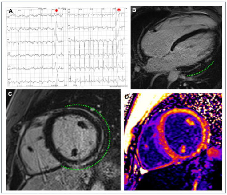
Figure 5.
Example of a myocardial scar in an athlete. A competitive hockey player aged 26 at the pre-participation screening during exercise testing showed frequent PVBs with right bundle branch block/superior axis morphology at high workload ((A), red asterisks). Post-contrast sequences on CMR revealed a subepicardial stria of LGE involving the anterior, lateral, and inferior LV walls in their basal and medium portions, with a “ring-like” pattern (green dotted line; (B) 4-chamber view; (C) short-axis view). Increased signal in the correspondent areas of fibrosis in the native T1 mapping short-axis sequence (D). Reproduced with permission from Brunetti et al. [129].

