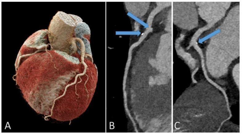
Figure 6.
Representative example of coronary calcifications in a 56-year-old endurance athlete without risk factors who underwent coronary computed tomography for investigation of repolarization abnormalities on resting ECG. 3D reconstruction of the coronary artery tree (A). Calcific plaques in left anterior descending (arrows, B) and circumflex (arrow, C) coronary arteries.

