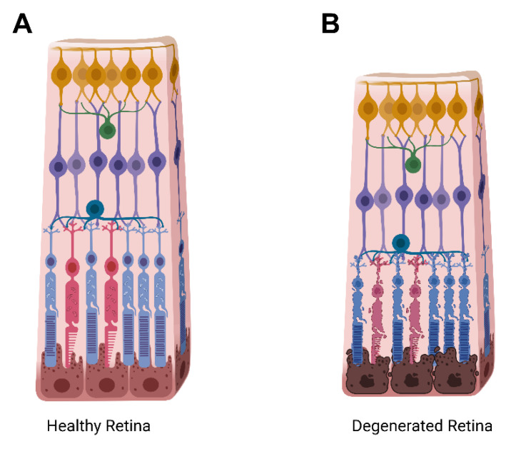
Figure 2.
Cartoon schematic of a healthy retina in comparison to a degenerating retina. (A) Illustration of the retinal layers in an intact, healthy, retina. (B) Illustration of a degenerating retina with rod photoreceptors depicted in blue, and cone photoreceptors depicted in red. The total retinal thickness, as well as the outer nuclear layer containing the rods and cones thins upon degeneration as photoreceptor cells shrink, lose functionality, and die. Yellow, ganglion cells; green, amacrine cells; teal, horizontal cells; purple, bipolar cells; brown, retinal pigment epithelium.

