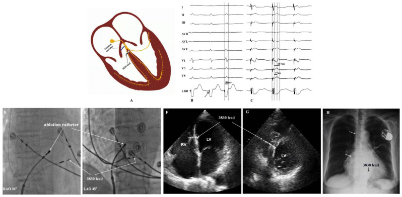
Figure 1.
Left bundle branch area pacing. (A): Diagram of the LBBaP and AVNA. (B): LBB potential recorded from the 3830 lead (dash arrow). Baseline QRSd of 88 ms. (C): The 3830 lead implanted in LBB area successfully; unipolar LBBaP pacing at pacing at 1 V/0.4 ms resulted in LBB capture with QRSd of 107 ms and Stim-LVAT of 77 ms. (D,E): Fluoroscopic imaging of LBBaP lead implantation ((D): RAO 30-deegree fluoroscopic views and (E): LAO 45-degree fluoroscopic view). (F,G): Echo images demonstrating the location of the LBBaP lead in the interventricular septum ((F): apical 4-chamber view and (G): left parasternal short axis transection view). (H): Radiograph of chest after LBBaP and AVN ablation (posteroanterior view). LBB = left bundle branch, LBBaP = left bundle branch area pacing, AVNA = atrioventricular node ablation, RAO = right anterior oblique, LAO = left anterior oblique, RV = right ventricle, LV = left ventricle, Stim-LVAT = pacing stimulus to left ventricular activation time, Echo = echocardiography.

