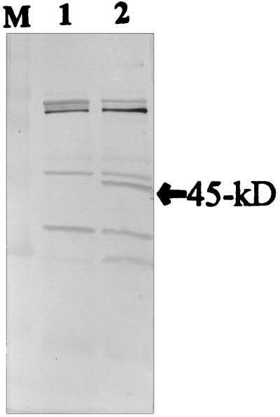
FIG. 1.
Immunoblot (Western blot) analysis with the polyclonal antibody raised against whole-cell proteins of S. suis type 2. Lanes: M, rainbow molecular weight marker (Amersham); 1, E. coli DH5α(pUC19) negative control; 2, E. coli DH5α(pOT401). The location of the 45-kDa GDH protein is indicated by the arrow.

