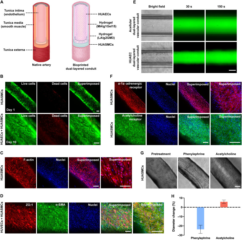
Fig. 4. Structural and biological functions of (bio)printed arterial conduits.
(A) Schematics showing structures of the native artery and (bio)printed arterial conduit. (B) Fluorescence microscopic images showing the viability of (bio)printed HUASMCs at different time points of culture. Green, live cells; red, dead cells. Scale bars, 100 μm. (C) Fluorescence confocal images of the immunostained artery exhibiting expressions of F-actin by HUASMCs. The cells were counterstained with DAPI for nuclei. Red, F-actin; blue, nuclei. Scale bars, 100 μm. (D) Fluorescence confocal images of the immunostained artery exhibiting expressions of ZO-1 by HUAECs and α-SMA by HUASMCs. The cells were counterstained with DAPI for nuclei. Red, ZO-1; green, α-SMA; blue, nuclei. Scale bars, 100 μm. (E) Diffusion of 3- to 5-kDa FITC-Dex in the (bio)printed dual-layered conduits in the absence (top) and presence (bottom) of endothelium formed by HUAECs in the lumens. Scale bar, 200 μm. (F) Fluorescence confocal images of the immunostained artery exhibiting expressions of α-1a-adrenergic receptor and muscarinic acetylcholine receptor by HUASMCs. The cells were counterstained with DAPI for nuclei. Red, α-1a-adrenergic receptor; green, muscarinic acetylcholine receptor; blue, nuclei. Scale bars, 100 μm. (G) Vasoactivities of bioprinted arterial conduits in response to phenylephrine and acetylcholine. Scale bar, 200 μm. (H) Quantified changes in diameter of bioprinted arterial conduits in response to phenylephrine and acetylcholine.

