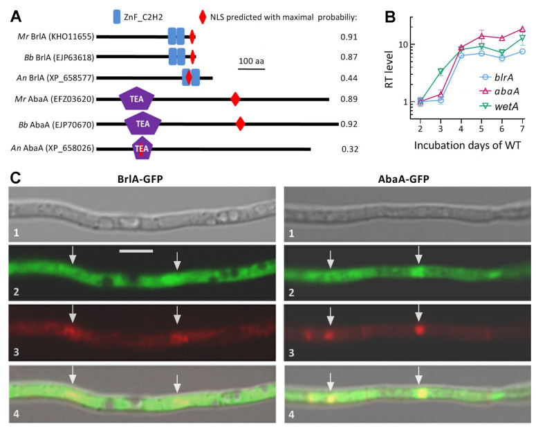
Figure 1.
Domain architectures, transcriptional profiles, and subcellular localization of BrlA and AbaA in M. robertsii (Mr). (A) Comparison of conserved domains and NLS motif predicted from amino acid sequences of BrlA and AbaA orthologs. An, A. nidulans. Bb, B. bassiana. (B) Relative transcript (RT) levels of three CDP genes in the Mr WT strain during a 7-day incubation on PDA at an optimal regime with respect to a standard on day 2. Error bars: standard deviations (SDs) of the means from three independent cDNA samples analyzed via qPCR. (C) LSCM images (scale bar: 5 μm) for subcellular localization of the BrlA-GFP and AbaA-GFP fusion proteins expressed in the WT strain. Images 1, 2, 3, and 4 are bright, expressed, DAPI-stained, and merged views of the same field, respectively. Hyphal nuclei are indicated by arrows.

