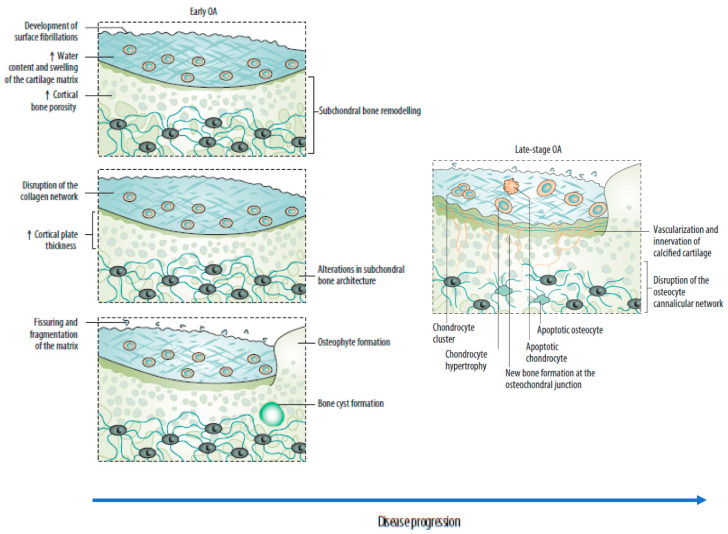
Figure 2.
Sequential changes in the osteochondral unit during the evolution of osteoarthritis. (Left) Early OA is characterized by increased remodeling of the subchondral bone plate. With disease progression, loss of cartilage matrix proteoglycans and erosion of the collagen network led to the development of deep fissures and delamination of the cartilage, with exposure of the underlying zones of calcified cartilage and subchondral bone. In the subchondral bone, cortical plate thickness gradually increases. (Right) Chondrocytes exist mostly in clusters in late-stage OA, but chondrocyte apoptosis is also evident. In the deeper zones, chondrocytes undergo phenotypic alterations, developing features of a hypertrophic phenotype. The calcified cartilage expands and advances into the overlying hyaline articular cartilage, with duplication of the tidemark. This process is initiated by penetrating vascular elements, and accompanying sensory and sympathetic nerves, into the osteochondral junction. OA: osteoarthritis. Source: original.

