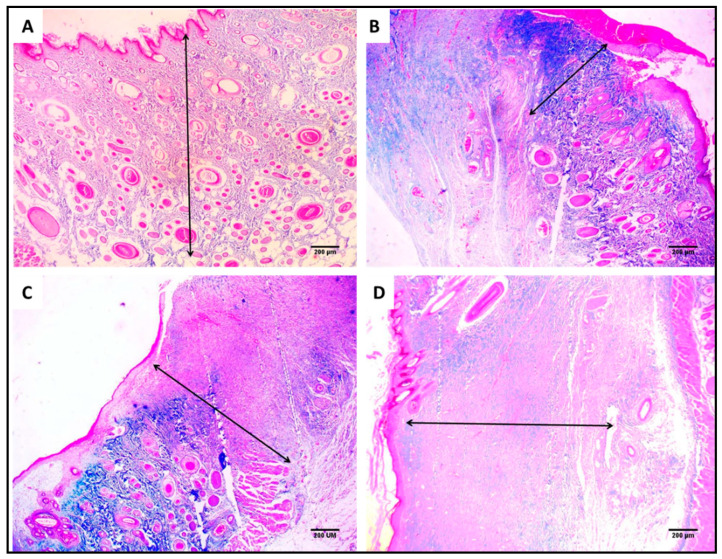
Figure 14.
Masson’s trichrome staining of the wound showing the area of collagen fibers (%) using image J software of (A) section in normal skin (negative control) showing 28.2% area of collagen fiber (×40). (B) Section in the wound of the control group showing 8.2% area of collagen fiber (×40). (C) Section in the Betadine™-treated group showing 14.7% area of collagen fiber (×40). (D) Section in the AgNPs-treated group showing 22.4% area of collagen fiber (×40).

