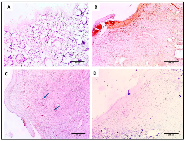
Figure 15.
TNF-α immunohistochemical staining of (A) section in normal skin (negative control) showing negative TNF-α immunostaining (−) as no wound healing area (×100). (B) Section in the wound of the control group showing strong positive immunostaining (+++) in the wound healing area (×100). (C) Section in the Betadine™-treated group showing mild positive TNF-α immunostaining (+) in the wound healing area (×100). (D) Section in the AgNPs-treated group showing negative TNF-α immunostaining (−) in the wound healing area (×100).

