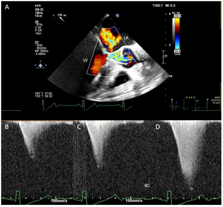
Figure 1.
(A) Transesophageal color Doppler method showing the systolic anterior motion of the mitral valve and aliased flow in the left ventricular outflow tract during systole. Continuous wave Doppler showed a gradient ranging between 50 (B), 80 (C), and 180 mmHg (D) in different steps of the surgical procedure. LA, left atrium; LV, left ventricle; AO, aorta.

