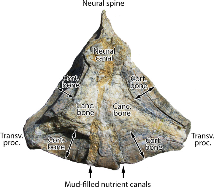
Fig 3. Transverse cross section through a caudal vertebra of Antaecetus aithai showing the dense, thickly-laminated, osteosclerotic cortical bone surrounding a central core of cancellous bone.
Cortical bone (arrows) is approximately 2 cm thick on the lateral sides of the centrum and 3 cm thick on the ventral side. Cancellous bone (white) is recrystallized, and the neural canal is now filled with crystalline matrix. Mud-filled nutrient canals are stained yellow. This half-centrum is approximately 14 cm wide, left to right, as shown. Specimen photographed in the field at Gueran.

