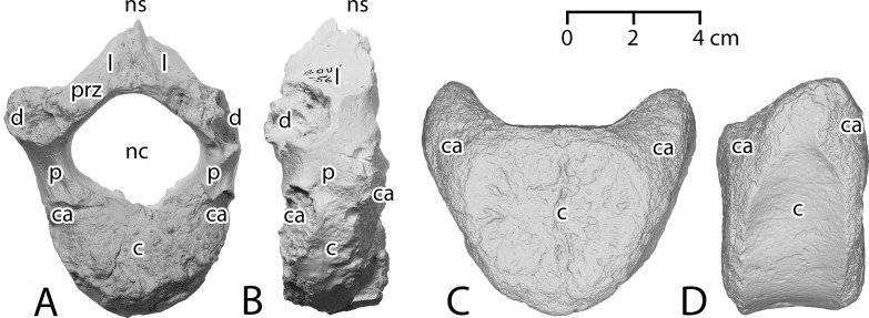
Fig 8. Anterior thoracic vertebrae of Antaecetus aithai and ‘Pachycetus’ humilis.
A, thoracic T1 of A. aithae, FSAC Bouj-56, in anterior view; note the reniform centrum and large neural canal. B, FSAC Bouj-56 in left lateral view. C, centrum of T3 or T4 of ‘P.’ humilis, MMGD NsT-94, in anterior view; note the larger size and more circular centrum. D, MMGD NsT-94 in left lateral view. Illustrations A–B are photographs of a high-fidelity cast, and C–D show a high-resolution 3D digital scan, all reproduced at the same scale. Abbreviations: c, centrum; ca, capitular articulation; d, diapophysis; l, lamina; nc, neural canal; ns, neural spine; p, pedicle; prz, prezygapophysis.

