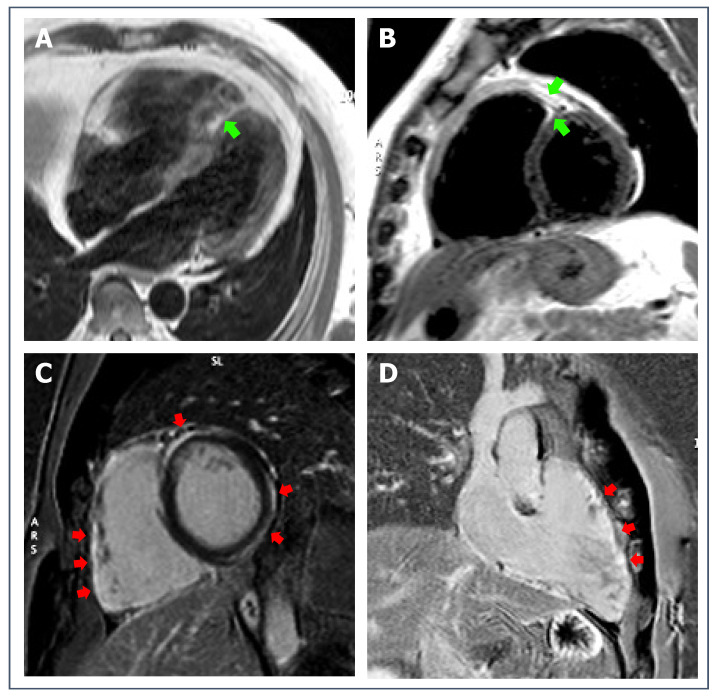
Figure 7.
43-year-old patient with biventricular ACM. T1-weighted sequences (A), 4-chambers view, and (B), short-axis view) show mild signs of fatty infiltration (green arrows). Post-contrast sequences (C), short-axis view, and (D), right heart 2-chambers view) reveal non-ischemic LGE involving both RV and LV walls (red arrows).

