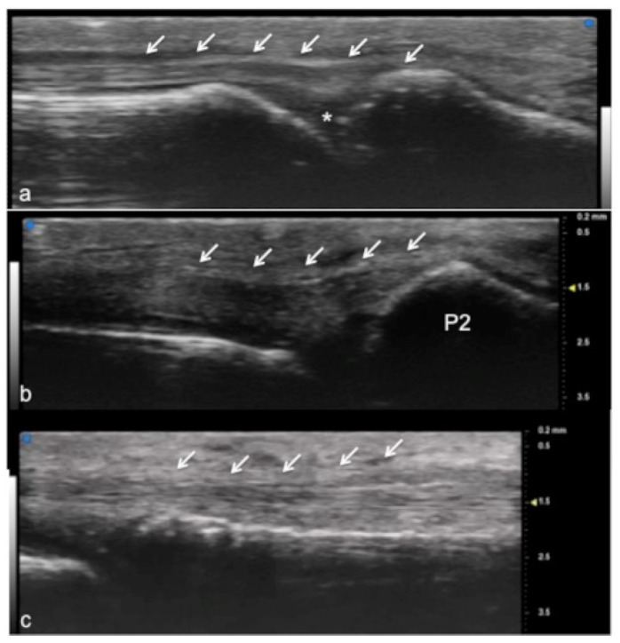
Figure 2.
Extensor tendon of a finger. UHFUS gives a detailed and magnified representation of small structures such as the extensor tendon of the finger, even allowing the visualization of partial lesions. In (a), the sagittal view of the terminal extensor tendon (white arrows) at the level of the distal interphalangeal joint (white asterisk). In (b), the median band of the extensor tendon inserting at the level of the middle phalanx (P2). In (c), the thin sagittal band of the extensor tendon at the level of the metacarpal head.

