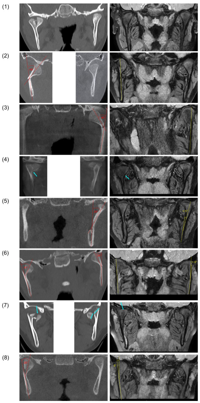
Figure 3.
Pretreatment CBCT/CT images (left) and follow-up magnetic resonance images (right) after fractures of the mandibular condyle and FOT in eight patients (rows 1–8). Angulation between fracture fragments is indicated in red (pretreatment) and yellow (follow-up). Fracture lines and bony scar lesions are marked by blue arrows.

