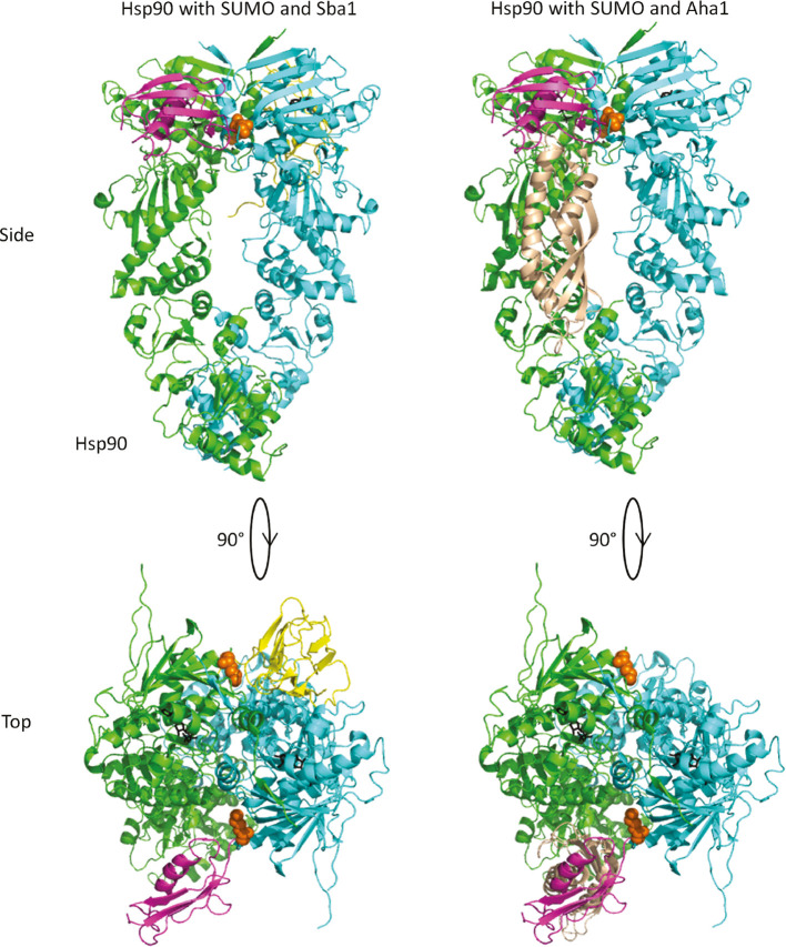
Figure 7:
A model for Sba1p and Aha1p binding to SUMOylated Hsp82p.
The Hsp82p dimer is shown in green and cyan. Lysine 178 is shown in orange spheres. Sba1p and Aha1p (N-domain only from PDBID 1USV; Meyer et al., 2004) are shown in yellow and beige, respectively. Smt3p is shown in magenta and modeled near lysine 178 (Sheng and Liao, 2002). The left panel shows that Sba1p and Smt3p coupled to position 178 of Hsp82p could occupy a similar space (each shown on the opposite side of the dimer). The right panel shows the position of the Aha1p N domain near where Smt3p would be coupled to lysine 178. PDBIDs 2CG9, 1L2N, 1USV (Sheng and Liao, 2002; Meyer et al., 2004; Ali et al., 2006).

