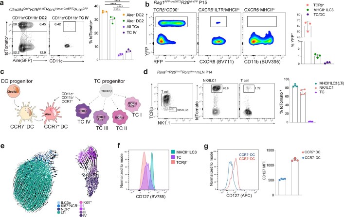
Extended Data Fig. 6. Thetis cells are ontogenically distinct from dendritic cells and ILC3s.
a, tdTomato labeling in cDC and TC from mLN of DC fate-mapping RORγt and Aire double reporter (Clec9aCre/+R26lsl-tdTomatoRorcVenus-creERT2AireGFP) mice at P18. b, Flow cytometry analysis of TCRβ+, MHCII+ ILC3, and CXCR6–MHCII+ cells encompassing TCs and DCs, from mLN of RAG1 fate-mapped (Rag1creERT2R26lsl-YFP) mice (n = 3) at P15 following 4-OHT treatment on P3, 5 and 7. c, Schematic of DC and TC ontogeny demonstrating distinct and overlapping transcriptional regulators and cell surface markers. d, Flow cytometry analysis of indicated immune cell subsets from mLN of RORα fate-mapped RorcVenus mice and summary bar graph for tdTomato labeling. e, UMAP of RORγt+MHCII+ cells (Fig. 2b) with scVelo-projected velocities, shown as streamlines. f, IL7R (CD127) expression (representative of n = 4 mice) on ILC3s and TCs. g, Expression of IL7R by DC subsets. Each dot represents an individual mouse, (n = 4). Data in a,b,d,f,g are representative of 2-3 independent experiments.

