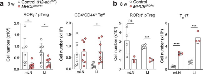
Extended Data Fig. 1. Analysis of pTreg cell generation in mice harboring MHC class II-deficient RORγt+ APCs.
a, Quantification of total pTreg (RORγt+Foxp3+) and CD4+ Teff (Foxp3–CD44hi) cells in the mesenteric lymph nodes (mLN) and large intestine lamina propria (LI) of 3-week-old MHCIIΔRORγt and control (H2-Ab1fl/fl) mice (n = 7 or 8 mice per group). b, pTreg (RORγt+Foxp3+) and TH17 (Foxp3–CD44hiRORγt+) cells in mLN and LI of 8-week-old MHCIIΔRORγt (n = 4) and control (n = 3) mice. Data in a pooled from two independent experiments. Data in b representative of three independent experiments. Error bars: means ± s.e.m. Statistics were calculated by unpaired two-sided t-test; *P < 0.05; ***P < 0.01, ****P < 0.0001.

