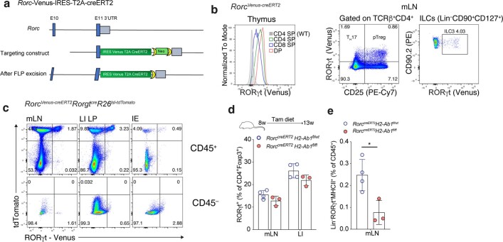
Extended Data Fig. 2. Temporal ablation of MHC Class II on RORγt+ APCs.
a, Targeting strategy for the Rorc locus. b, Flow cytometry of Venus expression in thymocytes (left) or mLN TCRβ+CD4+ T cells (middle) and Lin–CD90+CD127+ innate lymphoid cells (ILC; right) isolated from adult mice. c, Flow cytometry of mLN, LI and intestinal epithelial (IE) CD45+ and CD45– cells in P16 Rorc reporter RORγt fate-mapper (RorcVenus-creERT2RorgtcreRosa26lsl-tdTomato) mice. Representative of n = 3 mice. d-e, Frequency of pTreg cells amongst CD4+Foxp3+ cells in mLN and LI (d) or frequency of RORγt+ APCs (Lin–RORγt(Venus)+MHCII+) (e) in mLN of RorcVenus-creERT2H2-Ab1fl/fl (n = 4) or RorcVenus-creERT2H2-Ab1fl/wt (n = 3) mice maintained on tamoxifen diet from 8–13 weeks of age. Each symbol represents an individual mouse. Data in b–d representative of two independent experiments. Error bars: means ± s.e.m.; *P < 0.05; unpaired two-sided t-test.

