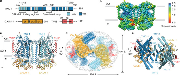
Fig. 1. Architecture and subunit arrangement of the TMC-1 complex.
a, Schematic representation of protein constructs that co-purified with TMC-1. EF1–EF3, EF-hand domains; TM, transmembrane domain. b, Local resolution map of the native TMC-1 complex after 3D reconstruction. c, Overall architecture of the native TMC-1 complex, viewed parallel to the membrane. TMC-1 (dark blue and light blue), CALM-1 (orange and yellow) and TMIE (red and pink) are shown in a cartoon diagram. Lipid-like molecules, N-glycans and putative ions are coloured black, green, and dark red, respectively. d, Cytosolic view of the reconstructed map fit to the model. Subunit densities are coloured as in c and the detergent micelle is shown in grey. e, A top-down extracellular view of the TMC-1 complex shows the domain-swapped dimeric interface. α-helices are represented as cylinders.

