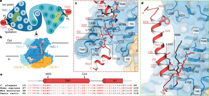
Fig. 2. TMIE resides on the periphery of the TMC-1 complex.
a, Schematic representation of TMC-1 (blue) and TMIE (red) transmembrane helices highlights the proximity of TMIE to the putative TMC-1 ion-conduction pathway. Palmitoylation of TMIE C44 and phospholipids (PL) is shown in black. b, Overview of the interaction interface between TMIE and TMC-1, viewed from the side. c, The interface between the TMIE ‘elbow’ and TMC-1. Interacting residues are shown as sticks. d, The interface between TMIE transmembrane helix and TMC-1, highlighting key residues and lipids. Palmitoylation is shown in red and phospholipid is shown in black. e, Multiple sequence alignment of TMIE orthologues. Elements of secondary structure are shown above the sequences and key residues are indicated with black arrows. Residues in black are not conserved, those in red are conservatively substituted, and those in bold red are conserved.

