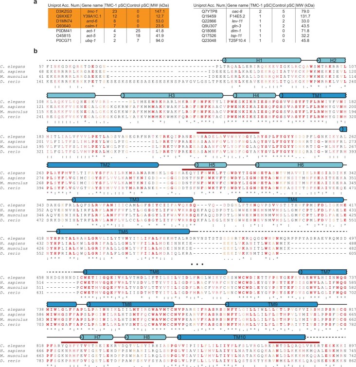
Extended Data Fig. 2. MS analysis of the TMC-1 complex.
a, Proteins detected by MS, via their associated peptide fragments, are listed with their gene name and molecular mass. The number of identified unique peptides from both the native TMC-1 complex and from wild-type worms (C. elegans N2), used as a control, are also indicated. b, Amino acid sequence and secondary structure of C. elegans TMC-1 are shown. The secondary structure based on the cryo-EM structure is indicated above the sequences as cylinders (α-helices), black lines (loop regions), or dashed lines (disordered residues). Red lines above the sequences indicate C. elegans peptides found by MS. . Note that the TMC-1 segments, corresponding to the sequence of 13–33, 557–566, 567–587, 877–890, 897–904, 917–927, 972–996, 1041–1052, 1177–1190, 1192–1216, and 1261–1269 are also found by MS, but not indicated in b.

