Fig. 5.
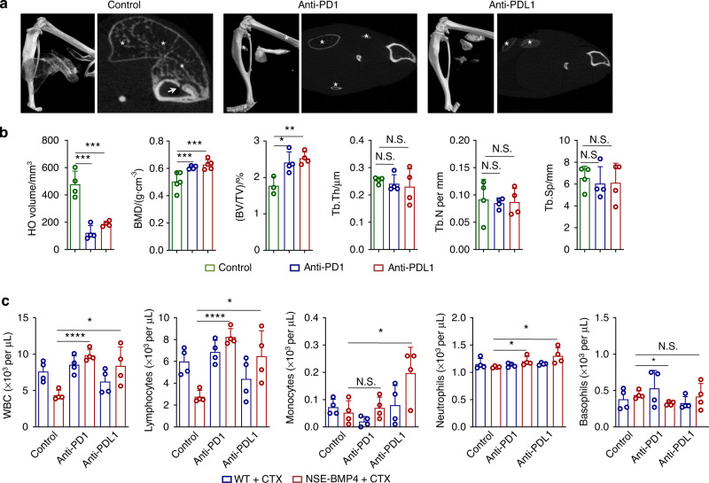
Neutralization of PD1/PDL1 alleviates HO progression and bone loss. a Representative microCT images of injured hindlimbs and a selected transverse section of tibia in the NSE-BMP4 mice treated with or without immune checkpoint blockade for two weeks. Arrows indicate bone mass loss. Asterisks indicate HO. b Statistical analysis of the HO volume, BMD and other bone parameters in the injured NSE-BMP4 mice following treatment with anti-PD1 or anti-PDL1 Abs (n = 4-5 per group). Data are presented as the mean ± s.d. of biological replicates. *P < 0.05, ***P < 0.001. N.S. indicates no significance (unpaired two-tailed t test). c Statistical analysis of the numbers of peripheral WBCs, lymphocytes, monocytes, neutrophils and basophils in the injured WT and NSE-BMP4 mice following PD1 or PDL1 blockade (n = 3 per group). Data are presented as the mean ± s.d. of biological replicates. *P < 0.05, ****P < 0.000 1. N.S. indicates no significance (unpaired two-tailed t test)

