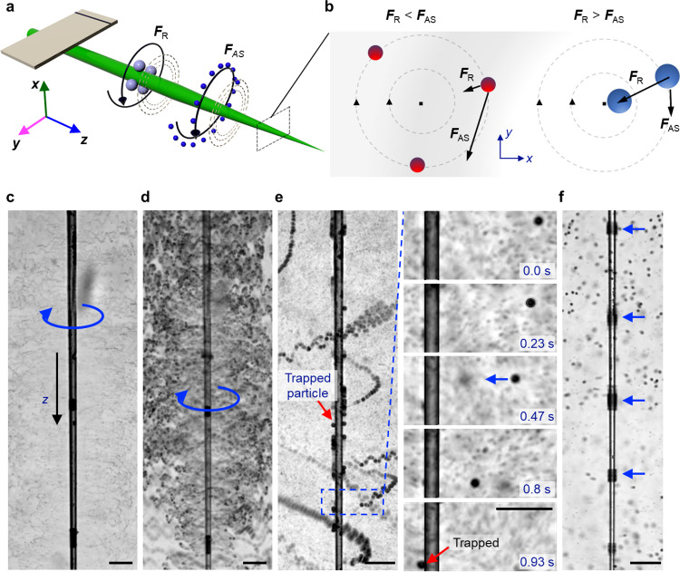
Fig. 5. Interaction and selective trapping of microparticles in an acoustically activated glass capillary.
a Schematic illustrating the selective trapping of microparticles along the acoustofluidic capillary system. Large particles, illustrated in red, relocate and get trapped along the shaft of the glass capillary due to the radiation force (FR). Small particles, shown in blue, follow out-of-plane circular streaming (FAS) around the capillary shaft. b (Left panel) The acoustic streaming force dominates for particles with diameter . (Right panel) The radiation force dominates for particles with diameter . (blue arrow represents the direction of the movement of microparticles) c, d Image sequence demonstrating circular streaming of 2 and 10 µm polystyrene microparticles around a glass capillary at an excitation voltage of 100 kHz and amplitude 3 VPP (blue arrow represents the direction of the movement of microparticles and red arrow shows the trapped particles) (see Supplementary Movie 7). e Superimposed time-lapse images illustrating trapping of polystyrene microparticles along the shaft of a glass capillary at 120 kHz and 10 VPP. The image series zooms in on the area marked with a blue box and shows the trapping of a single 15 µm microparticle. f Trapping of 10 µm beads at the pressure nodes of a glass capillary at an excitation frequency of 270 kHz and amplitude 10 VPP (see Supplementary Movie 7) (here the blue arrow shows the location of particles). All scale bars: 100 µm.

