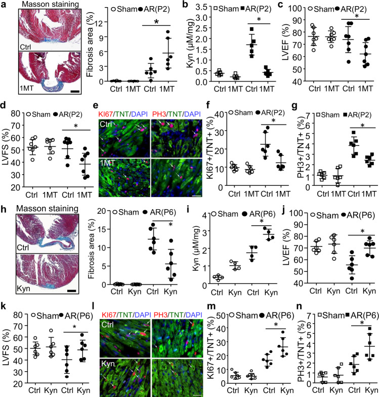
Fig. 2. The effects of Kyn on neonatal heart regeneration.
a–g Wild-type mice were intraperitoneally injected with IDO1 inhibitor (1-methyl tryptophan, 1MT, 100 mg/kg) or PBS (Control, Ctrl) every other day from P1 up to P28 and underwent heart AR surgery at P2. Cardiac fibrosis, function, and Kyn concentration were analyzed at P28. a Masson staining was used for detection and quantitation of fibrosis area (n = 6/group, *P = 0.0074). b Cardiac Kyn concentration was measured by HPLC (n = 5/group, *P < 0.0001). c, d Left ventricular ejection fraction (LVEF, *P = 0.0363) and left ventricular fraction section (LVFS, *P = 0.0156) were determined by echocardiogram (n = 7/group). e–g The cardiomyocyte proliferation was determined and quantified by co-staining of KI67 (*P = 0.0017) or PH3 (*P = 0.0031) with Troponin T (TNT) at P7 (n = 6/group). h–n Wild-type mice were intraperitoneally injected with PBS (Control, Ctrl) or Kyn (100 mg/kg) every other day from P1 up to P28 and underwent heart AR surgery at P6. Cardiac fibrosis, function, and Kyn concentration were analyzed at P28 (n = 6/group). The quantification of fibrosis area (h, n = 6/group, *P = 0.0007), cardiac Kyn concentration (i, n = 4/group, *P = 0.0007), Left ventricular ejection fraction (LVEF, j, *P = 0.0045) and left ventricular fraction section (LVFS, k, *P = 0.087) (n = 6/group), and cardiomyocyte proliferation (P10, l–n, n = 6/group, *P = 0.0048 for KI67, *P = 0.0095 for PH3) were analyzed. Bar = 500 µm for (a, h). Bar = 50 µm for e and l. Values are presented as means ± SD. *P < 0.05. Statistical analysis: one-way ANOVA followed by Tukey’s multiple comparisons test in a–h.

