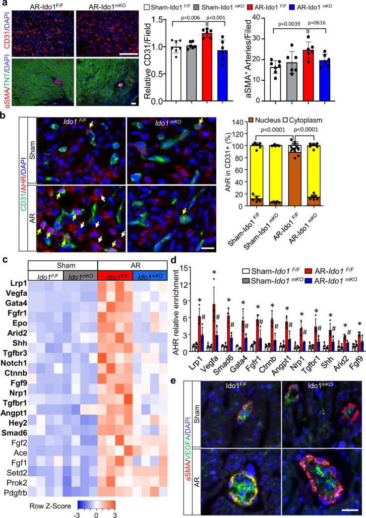
Fig. 4. Kyn-activated AhR promotes cardiac angiogenesis in cardiac regeneration.
Ido1 F/F (Ido1 Flox/Flox) and Ido1 mKO (Tnt Cre; Ido1 Flox/Flox) mice underwent heart AR or Sham surgery at P1 and were collected at P7 for cardiac angiogenesis study. a Representative images and quantification of CD31 (endothelial cell-specific marker) and α-SMA (smooth muscle cell-specific marker) immunofluorescence (IF) staining (n = 7 for Sham-Ido1 F/F group; n = 6 for other three groups). Bar = 100 µm. b Co-IF staining and quantification of the location of AHR at cytoplasm (white arrow) and nucleus (yellow arrow) in CD31-positive cells (n = 7 for Sham-Ido1 F/F group; n = 6 for other three groups). Bar = 20 µm. Two-way ANOVA followed by the Tukey’s test was used for statistical analysis. c Heatmap showing all significant changes (22/32) of the gene related to coronary vasculature morphogenesis in endothelial cells (ECs) isolated from apical resected heart compared with Sham hearts in Ido1 F/F mice. Genes with bold font indicate a significant decrease in resected Ido1 mKO vs. Ido1 F/F mice (n = 4/group). d Chromatin immunoprecipitation quantitative PCR (ChIP-qPCR) analysis of the enrichment of AHR for the indicated genes in ECs isolated from resected or Sham hearts (n = 4/group). e Representative co-IF staining of α-SMA and VEGFA for six mice of each group and five images of each mouse were collected. Bar = 20 µm. Data are mean ± SD. *P < 0.05 vs. Sham-Ido1 F/F group, # P < 0.05 vs. AR-Ido1 F/F group in (d). The Student t test (two-tailed) was used in (a, c, d).

