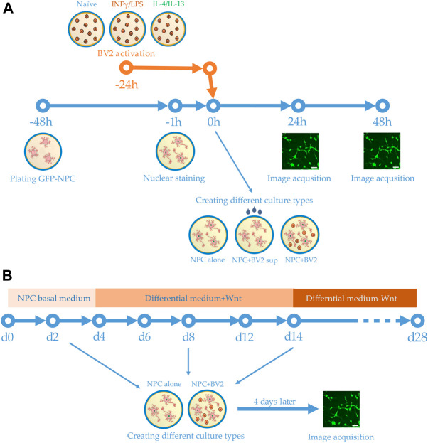
FIGURE 1.
Schemes of experimental design. (A) Human GFP-NPCs were plated 2 days prior to the experiments. Meanwhile BV2 cells were stimulated with proinflammatory agents or anti-inflammatory cytokines. One hour before the experiments, the nuclei of GFP-NPCs were stained. GFP-NPCs were exposed to microglia-conditioned media (sup) or co-cultured with microglia. GFP-NPCs alone served as control. Images were acquired using HCS at 0, 24, and 48 h. (B) Schematic representation of the differentiation protocol. Human NPCs were cultured in NPC basal medium for 4 days, then further cultured for an additional 10 days in differentiation media. After 2 weeks, the Wnt was omitted from the complemented media. Co-cultures were assembled on days 2, 8, and 14; immunofluorescence staining and image acquisition were performed after 4 days of co-culturing (on days 6, 12, and 18).

