FIGURE 3.
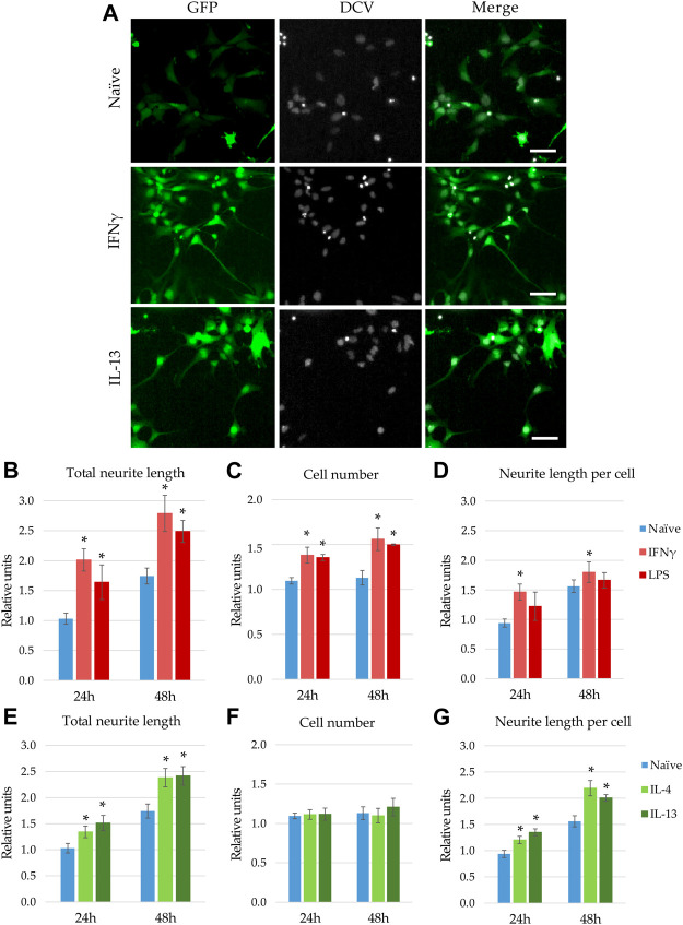
Modulatory effect of microglia stimulation on microglia-NPC interaction. GFP-NPCs were subjected to supernatants from BV2 cells previously stimulated with proinflammatory agents (IFNγ or LPS), or anti-inflammatory cytokines (IL-4 or IL-13). As controls, GFP-NPCs treated with supernatants from naïve BV2 cells were used. (A) Representative fluorescence images depict GFP fluorescence (green) and DCV counterstaining (grey) in GFP-NPCs exposed to naïve, IFNγ-, or IL-13-pretreated BV2 supernatants for 48 h. Scale bars: 25 µm. The total neurite lengths (B,E), the cell numbers (C,F), and the neurite lengths per cell (D,G) were determined at the indicated time points. The relative units represent the values normalized to time 0 points of untreated NPCs. Data are presented as mean ± SEM (n = 4–7). For statistical analysis, two-way ANOVA followed by Tukey’s HSD post hoc test was performed. Asterisks indicate significant differences as compared to treatment with naïve BV2 supernatants (p < 0.05).

