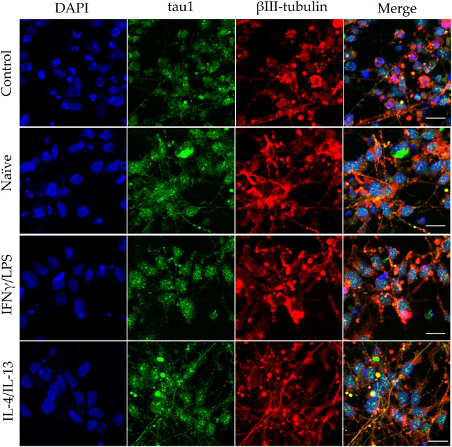
FIGURE 4.
Impact of naïve and stimulated microglia on the axon development of differentiating neurons. NPCs were differentiated toward the hippocampal DG granule cells for 18 days. The differentiating neuronal cells were co-cultured with BV2 cells for 4 days (from day 14 to day 18). Microglial cells remained untreated (naïve) or were previously stimulated with an IFNγ/LPS or an IL-4/IL-13 cocktail as indicated. Confocal images depict day 18, neuronal cultures immunostained for the axonal marker tau1 (green) and the neuronal marker βIII-tubulin (red). The samples were counterstained with DAPI (blue). Scale bars: 25 µm.

