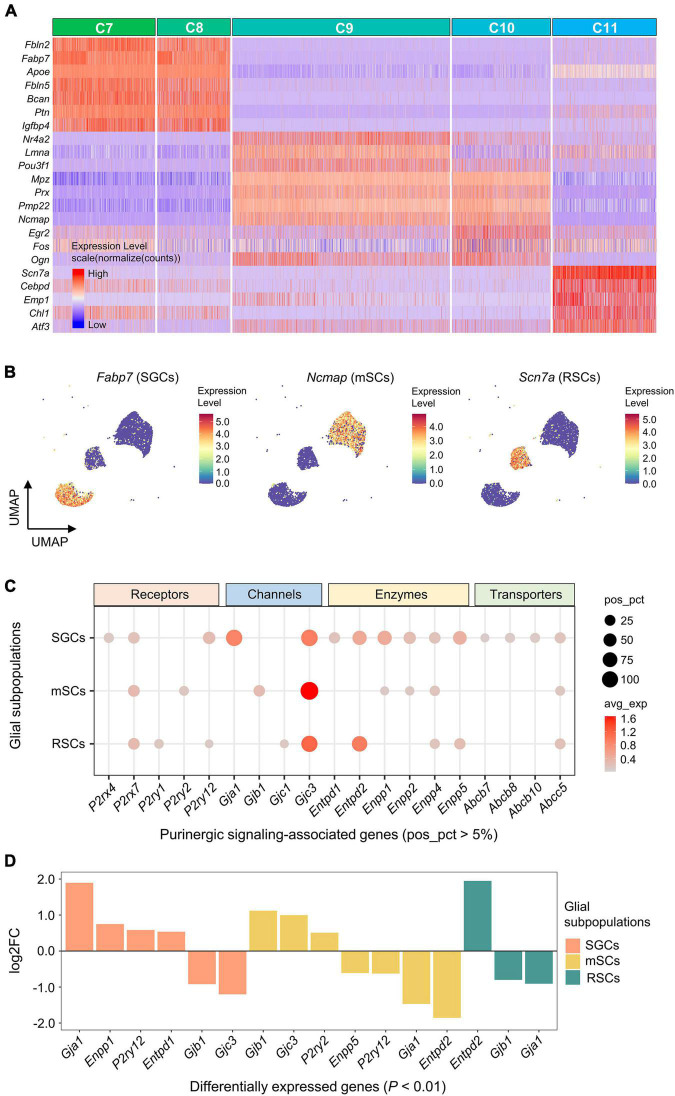
FIGURE 5.
Expression of genes associated with purinergic signaling in glial subpopulations. (A) Heatmap showing expression of representative marker genes for each glial cluster (C7–C11). Heatmap colors represent relative expression levels. Red represents high expression and blue low expression. Glial clusters were color-coded above the heatmap. (B) UMAP projection of known marker genes for SGCs, mSCs, and RSCs in glial clusters (C7–C11). Each dot represents one cell. Cells are colored by gene expression levels. Levels are normalized expression counts. Warm colors (red and yellow) represent high expression, and cool colors (light and dark blue) represent low expression. C7 and C8 were identified as SGCs by the expression of Fabp7, C9 and C10 were identified as mSCs by Ncmap, and C11 was identified as RSCs by Scn7a. (C) Bubble plot showing genes encoding purinergic complex (including receptors, channels, enzymes, and transporters) in glial subpopulations. (D) Bar plot showing differential expression of these genes among glial subpopulations (Wilcoxon Rank Sum test; P < 0.01 was considered statistically significant). The bars are color-coded by glial subpopulations, and bar height represents log2 fold change (log2FC). Genes with log2FC > 0.5 and P < 0.01 are shown. SGCs, satellite glial cells; mSCs, myelinating Schwann cells; RSCs, Remak Schwann cells.

