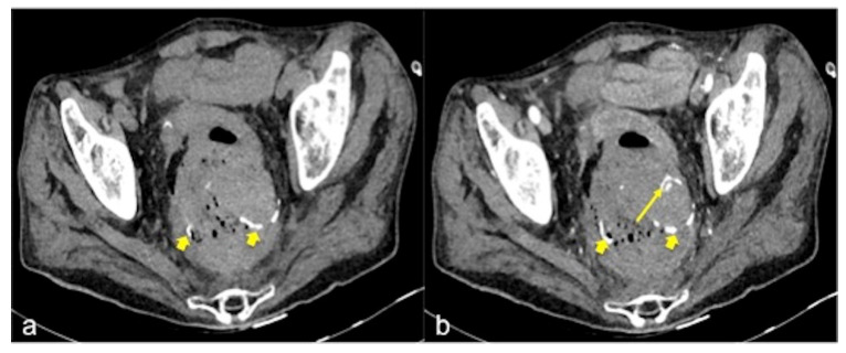
Figure 2.
CT multiphasic study of haemorrhage at the surgical site of colorectal anastomosis. The pre-contrast CT axial image (a) shows spontaneous hyperdensity of metallic clips (small arrow). The arterial phase (b) shows another hyperdense spot (long arrow) near the anastomosis and is suggestive of active bleeding.

