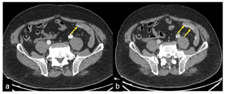
Figure 3.
CT multiphasic study of bleeding at the proximal tract of sigma. In the arterial axial image after contrast media administration (a) it is possible to detect a blush of active bleeding (arrow). In the venous phase (b) a dimensional increase of the hyperdense blush is present (arrows) as a sign of active incremental bleeding in the venous phase.

