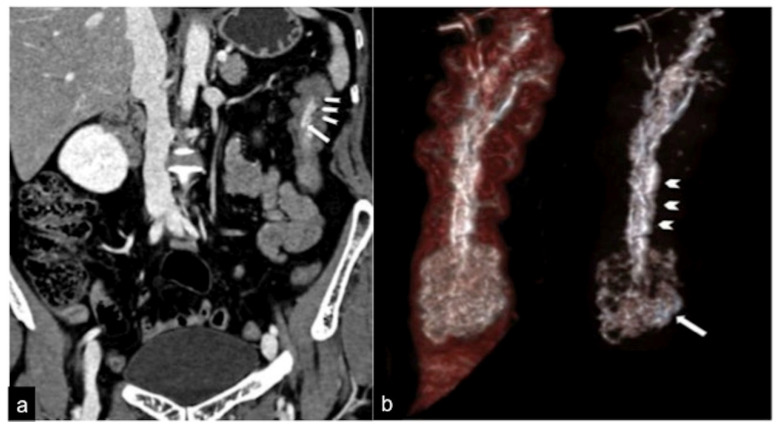
Figure 6.
CTA coronal reconstruction in a patient with sub-occlusive recurrent status and occult haemorrhage. The coronal reconstruction (a) detects a pedunculated polyp (long arrow) with an arterial axis of vascular support. In the MIP post-processing with MPR and VR-3D, the coronal view (b) better defines the polypoid formation and the axis of vascular support (head arrows). Adopted with permission from ref. [15].

