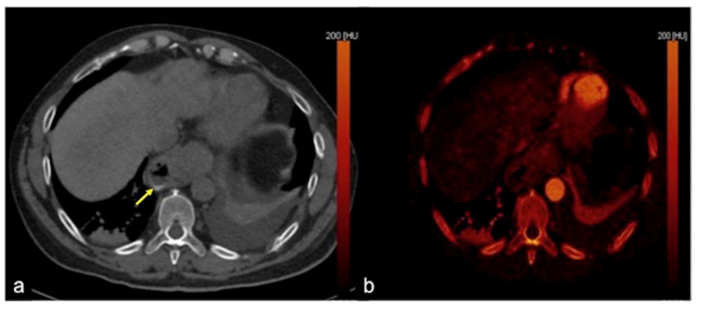
Figure 8.
DECTA multiphasic study reconstruction. The virtual non-contrast axial image (a) shows an endoluminal linear hyperdensity in the distal oesophagus (arrow). In the iodinated map axial image (b) the hyperdensity is not magnified. These findings are suggestive of the presence of a foreign body, confirming the high confidence of DECTA post-processing diagnostic performance.

