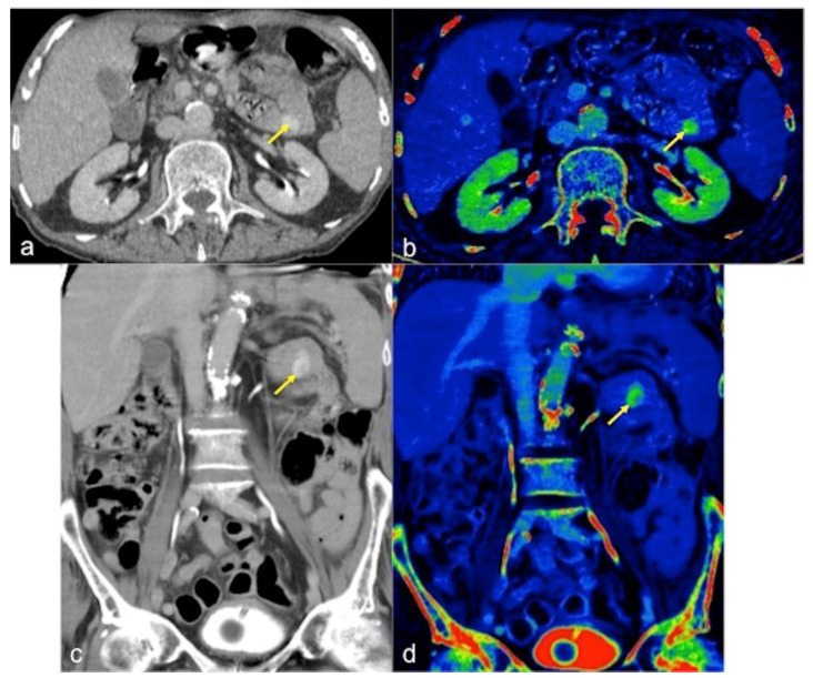
Figure 9.
DECTA multiphasic study of active bleeding in the proximal jejunum. Thanks to axial (a,b) and coronal (c,d) images, it is possible to easily compare the traditional greyscale and the colourimetric maps of iodium distribution; a haemorrhagic focus of active bleeding is detected with high sensitivity ((a–d) arrow).

