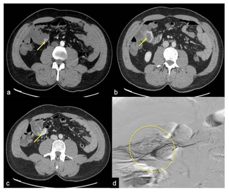
Figure 11.
CTA multiphasic study of small intestine angiodysplasia. In the arterial phase (a) an active blush of contrast media is detected at the small bowel wall (arrow). The venous (b) and late (c) phases show a gradual increase of contrast blush (arrow (b)) with consequent production of a haematic collection (arrow (c)). In the selective angiography (d) the CTA findings are confirmed and the presence of an angiodyslastic focus is documented (discontinued circle).

