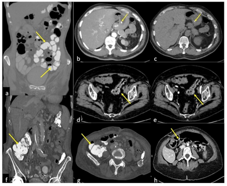
Figure 19.
Pitfalls in the detection of acute gastrointestinal bleeding. (a) Positive contrast media administration (arrow): coronal CTA reconstruction of contrast media distended bowel loops (arrows) obscure any GIB source. (b,c) Endoluminal hyperdense ingested capsule: arterial and pre-contrast axial CTA image of an endoluminal hyperdense ingested capsule in patients with melaena. In the arterial phase with MIP post-processing (b), an endoluminal hyperdense inclusion is detected (arrow) in the gastric lumen; the pre-contrast phase (c) confirms the presence of the inclusion with analogous morphologic and densitometric characteristics (arrow). (d,e) Retention of contrast media in patients with diverticulosis and haematochezia: pre-contrast (d) and arterial (e) axial CTA images show unchanged hyperdense diverticula (d,e, arrow). (f,g) Hyperdense fecaloma: coronal (f) and axial (g) CTA image of hyperdense fecaloma (f,g, arrow) due to retained contrast media. (h) Cone-beam artefacts: axial arterial CTA image shows massive hyperdensities within the bowel lumen (arrow) due to cone-beam artefacts.

