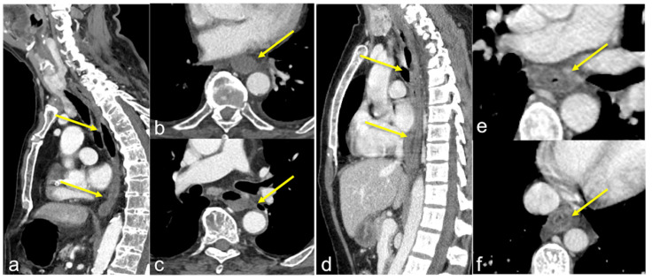
Figure 23.
Two different patients with consequences of caustic ingestion on the oesophagus. Enhanced CTA scan in the portal venous phase in sagittal (a,d) and axial (b,c,e,f) views. Note, in the first case (a–c) the extensive oesophageal necrosis (a–c, arrows) with a thin and unenhanced wall, in the second case (d–f) the oesophageal thickening with a stratified wall (d–f, arrows).

