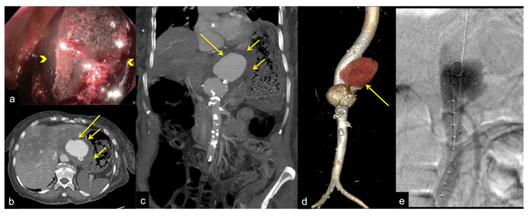
Figure 31.
Abdominal pain and anaemia. Gastric endoscopy (a) shows an extrinsic bulging mass in the gastric corpus with a large adherent blood clot ((a) arrowheads). Axial CTA artery phase (b) and coronal MIP artery phase reconstruction (c) show a contained rupture of a thoraco-abdominal aortic aneurysm ((b,c) long arrows) with periaortic haematoma compressing the posterior gastric wall with loss of interface fat plane ((b,c) short arrows). The VR-3D artery phase image reconstruction of the aortic aneurism (d, arrow) better defines the extension of the aneurysm sac ((d) long arrow) for endovascular operative planning. Angiography endovascular aneurysm repair procedure (e).

